Study Ecocardiography.
Assalam o alaikum.
Hello my steemit friends i hope you all are doing well. I have always had a deeps interest in heart related studies and how the humans heart works. As a futures cardiologist i loved learning about everything that helps us see and understand the heart better. One very important tool in cardiology is echocardiography. In this post i will share what i have learned about echocardiography and explain the five important views: parasternal long axis parasternal short axis apical four chambers subcostal view and suprasternal view. I hopes you will finds it interesting and easy to understand.
Echocardiography is a test thats uses sound waves to creates pictures of the heart. It helps doctors see the heart's size shape and movement. It also shows how well the heart is pumping blood. This test is safe and does not cause pain. We used a special machine called a probe or transducers to do it. The probe sends sounds waves into the body and collects the echo that bounces back. This echo creates the image of the heart.
Parasternal Long Axis View
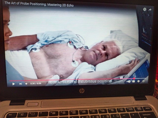
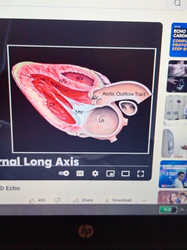
This is the first view we learn when starting echocardiography. In this view the probe is placed on the left side of the chest near the sternum (the breastbone). The patient usually lies on their left side. In this view we can see many important parts of the heart. We sees the left ventricle right ventricle left atrium aortic valve and mitral valve. This views helps us check the size of the heart chambers and how well the valves are working. It is a good view to detect valve problems like stenosis or regurgitation. This view is very helpful for beginners like me because it gives a clear picture of heart structure.
Parasternal Short Axis View.
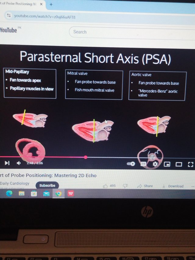
This view is taken by rotating the probe from the long axis view. The probe stays in the same place on the chest but we turn it 90 degrees. This gives us a circular or cross-sectional view of the heart. The parasternal short axis view lets us see the heart in slices like a cake. We can view the left ventricle at different levels – from the base (near the valves) to the middle (where the papillary muscles are) and to the apex (the tip of the heart). We also see the right ventricle and other structures. This view helps us understand how the heart contracts and how strong each part is working. It is very useful in patients with heart failure or muscle problems.
Apical Four Chambers View.
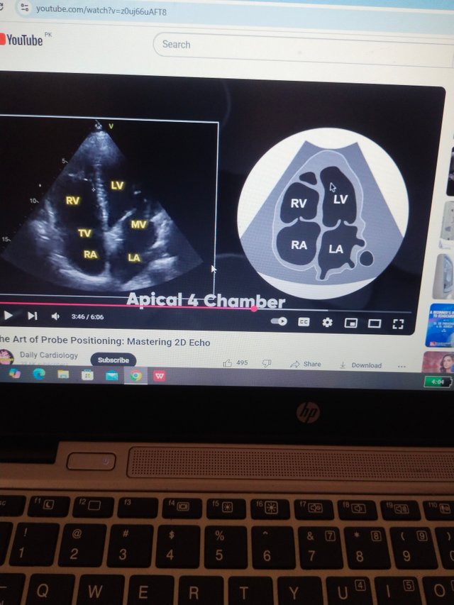
This is one of the most beautiful and complete views. The probe is placed at the apex of the heart which is usually around the fifth intercostal space at the midclavicular line. The patient lies on their left side for a better image. In this view we can see all four chambers of the heart clearly left atrium right atrium left ventricle and right ventricle. We also see the mitral and tricuspid valves. This view helps us compare the left and right sides of the heart. We can also check for valve leaks clots and congenital problems. It is very helpful in evaluating heart function. I love this view because it shows the heart working in real-time.
Subcostal View.
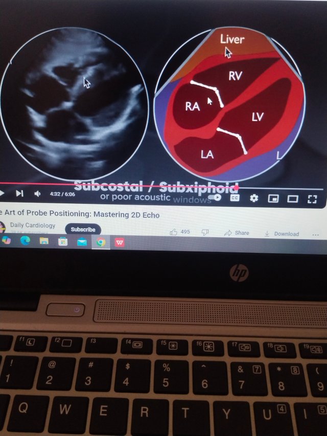
Sometimes we cannot get clear images from the chest especially in patients who are overweight have lung disease or are in critical condition. In such cases we use the subcostal view. For this view the probe is placed below the rib cage near the upper abdomen. The patient lies flat on their back. The sound waves go through the liver which helps us get a clearer picture of the heart. In the subcostal view we can see the four chambers of the heart like the apical view but from a different angle. It is also used to check for pericardial effusion (fluid around the heart). I think this view is very useful in emergency situations because its quick and easy to perform.
Suprasternal View.
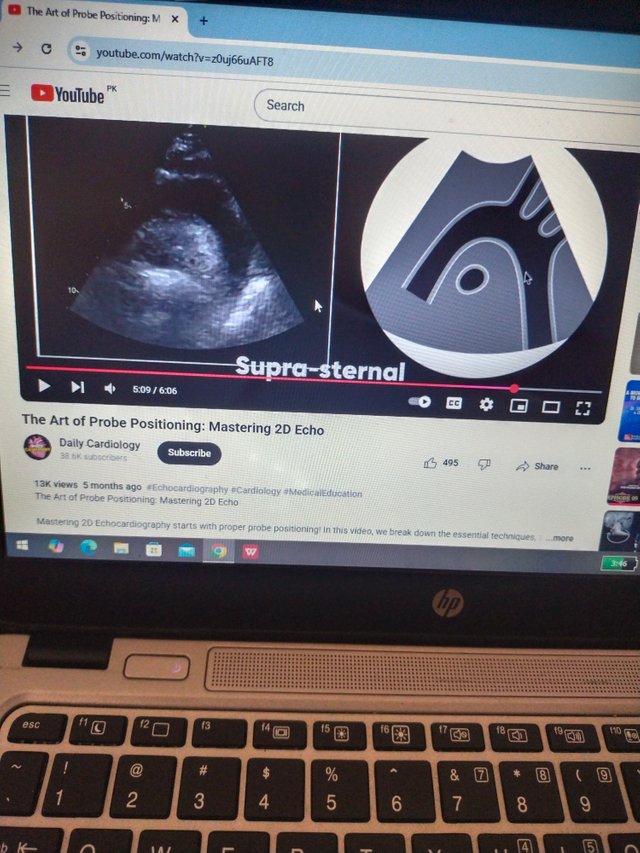
This is a special view that shows the large blood vessels coming from the heart like the aortic arch and its branches. The probe is placed above the sternum near the neck. In this view we can see the ascending aorta aortic arch and descending aorta. We can also see if there are any blockages aneurysms (bulges in the vessel) or dissection (a tear in the wall). This view is not used often but it is important for some heart conditions. It needs a good hand and patient cooperation so i am still practicing to become better at it.
Learning echocardiography is very exciting for me. These five views are the foundation for understanding the heart through ultrasound. Each views gives us different informations and together they helped doctors makes the right diagnosis. I feels proud that i am learning a skills that can save lives and help peoples in need. As I continued my studies i wants to become very skilled in using the echocardiography machines and reading heart images correctly.
Thank you for reading my post. I hope you enjoyed learning about the five views of echocardiography with me.
